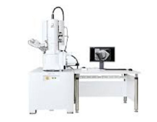
Since the invention of the microscope we have explored a world that was previously unimaginable. Everything that has been proven using this team has been the basis for many of the current advances. However since its creation, we have been looking for better and better ways to appreciate greater number of aspects of this world. That is why many varieties have emerged of microscopes. Discover the 5 advantages of the electron microscope over the conventional microscope.
What is the electron microscope? 🔬👨🔬
Built in 1931 by the German-born engineer, Ernst Ruska. A special kind of high-resolution microscope arose. Capable of enlarging the image of an object in nanometers, forming the same by using controlled electrons and a phosphorescent screen. In a short concept it can be defined as a microscope that uses an electron beam in an accelerated state as an illumination source.
Parts of the electron microscope 🔬🔌
Electron gun
It consists of a tungsten filament that when heated it generates electrons.


Electromagnetic lenses
In principle, before the impact on the sample there are two in total, the first condenser lens focuses the electron beam in the direction of the sample. On the other hand, the second lens organizes the electrons forming a narrow and thin beam.
After the incident on the sample is the lens objective which is located in the magnetic coils, has a great power and is where the first image is formed with an intermediate magnification.
Finally there is a third set of lenses magnetic lenses that are called projector lenses or eyepieces, which are they are responsible for the production of the final image with a large magnification.
Sample holder
It is a film of an extreme fineness that is formed by a metal grid and collodion or carbon.
Image display and recording system
It includes a fluorescent screen where the final image and a camera with which the image obtained can be recorded.
What is the fundamental principle of electron microscope? 🦠🧫
These devices use signals or reactions caused by the interaction between a sample and the incidence of an electron beam. Stop with this has information of its composition, morphology and structure.
Then we have that within its functioning it possesses essential parts such as the electron gun, which is the source of them.
Then it has two sets of condenser lenses whose function is to focus and to delimit the electrons in a narrow and thin beam. The displacement of the electrons through the column is employed voltages for acceleration between the anode and the tungsten filament.
The sample that are used in this type of microscopes it requires special preparation, more strict and delicate than conventional ones.
Taking into account the following:
It must be very thin, about two hundred times more than the one used in the optical microscope.Each sample to have some measures included between 20 and 100 nanometers and placed on the sample holders.
Then the electron beam passes through the sample and according to the refractive index of the different components and parts, they will disperse the electrons. Just below the sample is the objective lens. Which the remaining electron beam reaches and due to its high power is formed the image of intermediate size. Finally the ocular lens produces the image final well enlarged and detailed. Then the less dense areas will give rise to bright or clear regions within the image. While the dense areas by scattering more electrons, they will create darker areas in the final image.
Types of electron microscope 🔬🧬
The classification of the electron microscope is given due to its basic operating mechanism. Finding the following two:
Transmission electron microscope
Its main function is to analyze thin samples in such a way that electrons pass through them and generate an image by projection. It has many analogies compared to the traditional optical microscope. Among its most frequent uses we can find:
Obtaining images of the cellular interior.Obtaining structural images of the protein molecules, using metallic shading.Visualization of molecules in viruses by the negative staining technique.
Scanning electron microscope
This variety of electron microscope generates an image a from the emission of secondary electrons from the surface of the sample. In operation it is the analogue of optical stereo microscopes.
It receives this name since the image is formed with a kind of scanning performed with the electron beam in a raster pattern. Unlike the transmission it has sensors that capture the response of the electrons, thus forming the image.
Among the uses associated with this microscope we can find these:
Obtaining detailed images of the cell surface.Imaging of whole organisms.Particle counting and determination of its size.
Application 👩�🔬�


The main application of electron microscopy is the research deep understanding of the structure of molecules, micro-organisms, metals, cells and crystal. That is to conduct structure studies to a wide range of both inorganic and biological samples. In the industrial area it is used in the analysis of quality control failures.
One of its great applications is that they generate electronic microphotographs using specialized cameras. Is one of the fundamental pillars in the great advances of microbiology, since the structural study of bacteria and viruses has been of great help to the treatment.
Advantages of electron microscope
There are many advantages of its use, however we can to mention a few that are:
High image magnification.Amazing resolución.No there is an error due to distortion of the material during the preparation.Greater depth in the field of study.
Disadvantages of electron microscope
Live specimens cannot be studied.The sample preparation process is extended and thorough due to the thickness characteristics that are required.It must be worked with dried samples so that the preparation process is extensive.High cost maintenance.Your operators must have previous training for your use.
Now that you know the differences between the operation of the electron microscope and the light microscope. You can recognize the advantages granted by the use of electron microscopy in structural analysis. If you want to know about other microscopes, keep an eye on our upcoming publications.

